简介
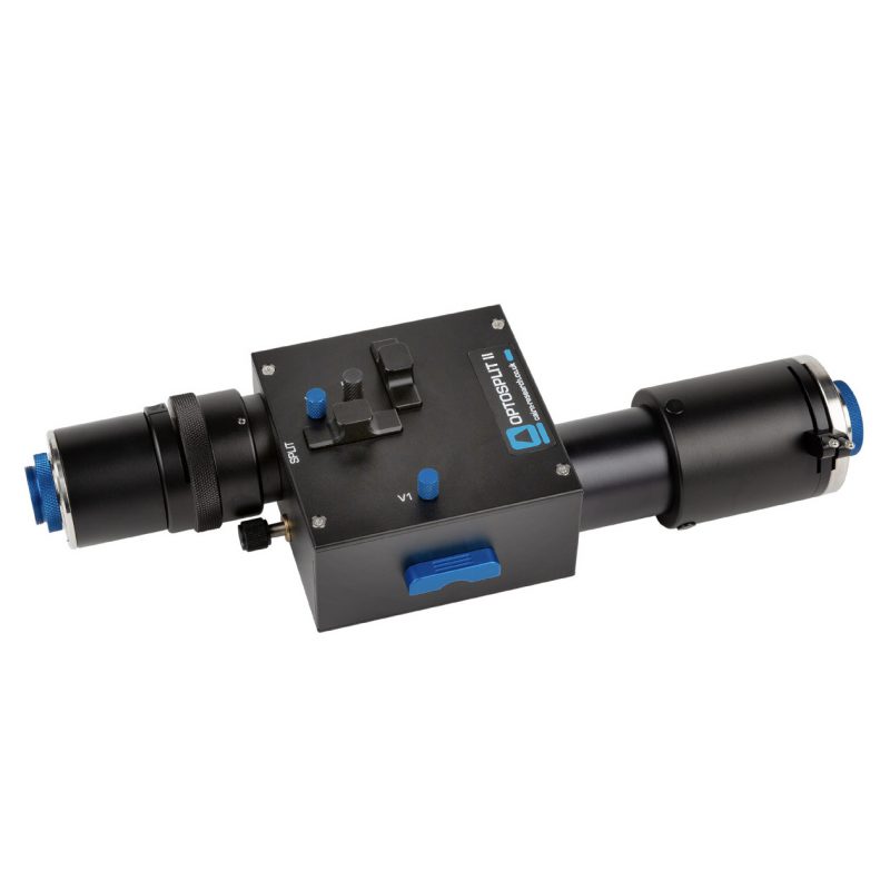
OptoSplit II 成像分离器是一台专业的光学分光仪器,可使单台相机同时记录两种不同波长、偏振状态或其他差异状态的图像。作为一种先进的荧光图像分离器,它满足了多样化的科研需求。
传统上,双通道成像是使用滤光片转换器或附加相机和分光器进行的,这两种方法都不是所有应用的理想选择。滤光片转换器的切换速度限制了时间分辨率,而第二台相机则增加了成本和复杂性。
OptoSplit 使用独特的旋转镜架,提供可调节的空间分离,以确保出色的图像配准,并具有完全可调的矩形光圈,可实现裁剪传感器成像模式并减少散射。
OptoSplit 采用特殊镜头设计,支持对角线长达 29.4 mm的传感器。它们具有相应更大的光圈和改进的离轴校正,可增强所有传感器的性能。作为分光装置的性能体现,大多数商业成像软件中都包含设备驱动程序,以协助配准并允许实时和离线配比或荧光叠加。
另外,OptoSplit 可以与其它图像采集软件一起使用,并手动离线进行处理,或者使用我们的 MicroManager 和 ImageJ 驱动程序。简单易用的设计使 OptoSplit II 成为多种应用的绝佳平台,例如,如双偏振成像;同时,适合用作同时成像装置。
虽然针对商用显微镜的耦合进行了优化,但成像分离器也可以与相机镜头或任何其他合适镜头系统一起使用。


应用
- 福斯特共振能量转移 (FRET)
- 比率钙、电压和 pH 成像
- 同时多荧光探针成像
- 偏振研究(各向异性)
- 同时相位对比/DIC 和荧光
- 同时双 Z 深度成像
- 全内反射荧光 (TIRF)
- 旋转盘共焦
- 单平面照明显微镜 (SPIM)
- 3D 超分辨率 PALM/STORM(使用柱面透镜)
特点
- 紧凑设计,标配集成 C 型接口输入和输出端口(可应要求提供 F 和 T 接口)
- 支持对角线长度高达 29.4mm的传感器
- 所有光学表面均采用 425nm 至 875nm AR 涂层,提升荧光分光器件性能
- 40mm 直径专有光学元件
- 简单而精确的图像配准控制
- 可互换滤光片/二向色镜支架,适合多种应用需求
相关文章
- IKCa channels control breast cancer metabolism including AMPK-driven autophagy
- Imaging Insulin Granule Dynamics in Human Pancreatic β-Cells Using Total Internal Reflection Fluorescence (TIRF) Microscopy
- Water cycles in a Hadean CO2 atmosphere drive the evolution of long DNA
- Escherichia coli chemotaxis is information limited
- Single-molecule imaging reveals replication fork coupled formation of G-quadruplex structures hinders local replication stress signaling
- Simultaneous spatiotemporal super-resolution and multi-parametric fluorescence microscopy
- An integrated multi-wavelength SCATTIRSTORM microscope combining TIRFM and IRM modalities for imaging cellulases and other processive enzymes
- E. coli chemotaxis is information-limited
- Quantitative fluorescence microscopy techniques to study three-dimensional organisation of T-cell signalling molecules
- A Multimodal Platform for Simultaneous T-cell Imaging, Defined Activation, and Mechanobiological Characterization
- Quantitative Intracellular pH Determinations in Single Live Mammalian Spermatozoa Using the Ratiometric Dye SNARF-5F
- Opposing modulation of Cx26 gap junctions and hemichannels by CO2
- Endoplasmic Reticulum Lumenal Indicators in Drosophila Reveal Effects of HSP-Related Mutations on Endoplasmic Reticulum Calcium Dynamics
- Optically activated, customizable, excitable cells
- Hot-Band Anti-Stokes Fluorescence Properties of Alexa Fluor 568


